Cause Alfalfa mosaic virus (AMV), which is transmitted by aphids, in seed, through pollen, and mechanically. The pea aphid, Acyrthosiphon pisum, is the main vector, but many other aphid species can transmit the virus.
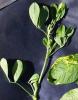
Note the yellow and green coloration of some of the leaflets.
Photo by C. M. Ocamb, 1998
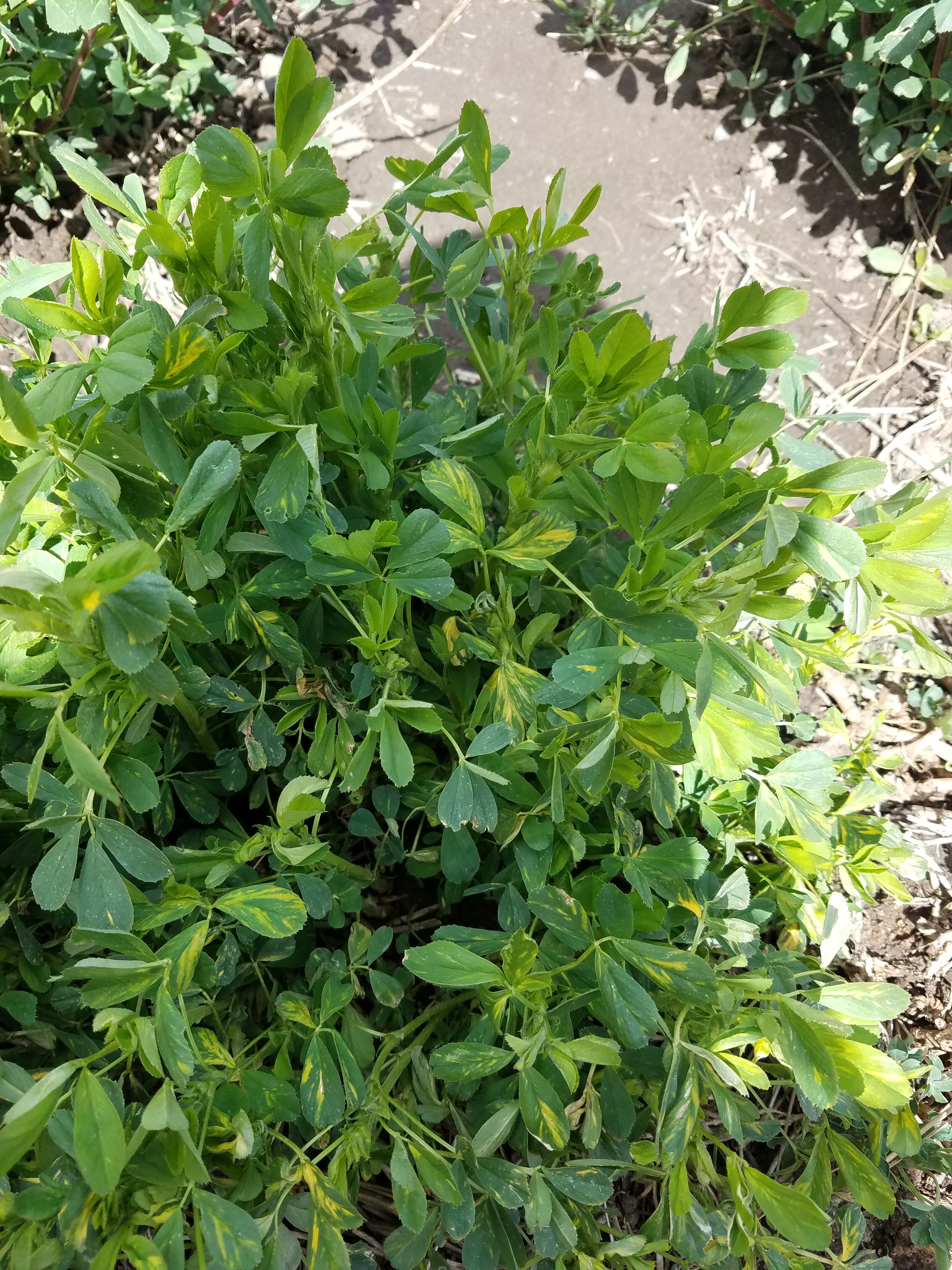
An alfalfa plant showing symptoms of AMV.
Photo by Ken Frost, 2020.
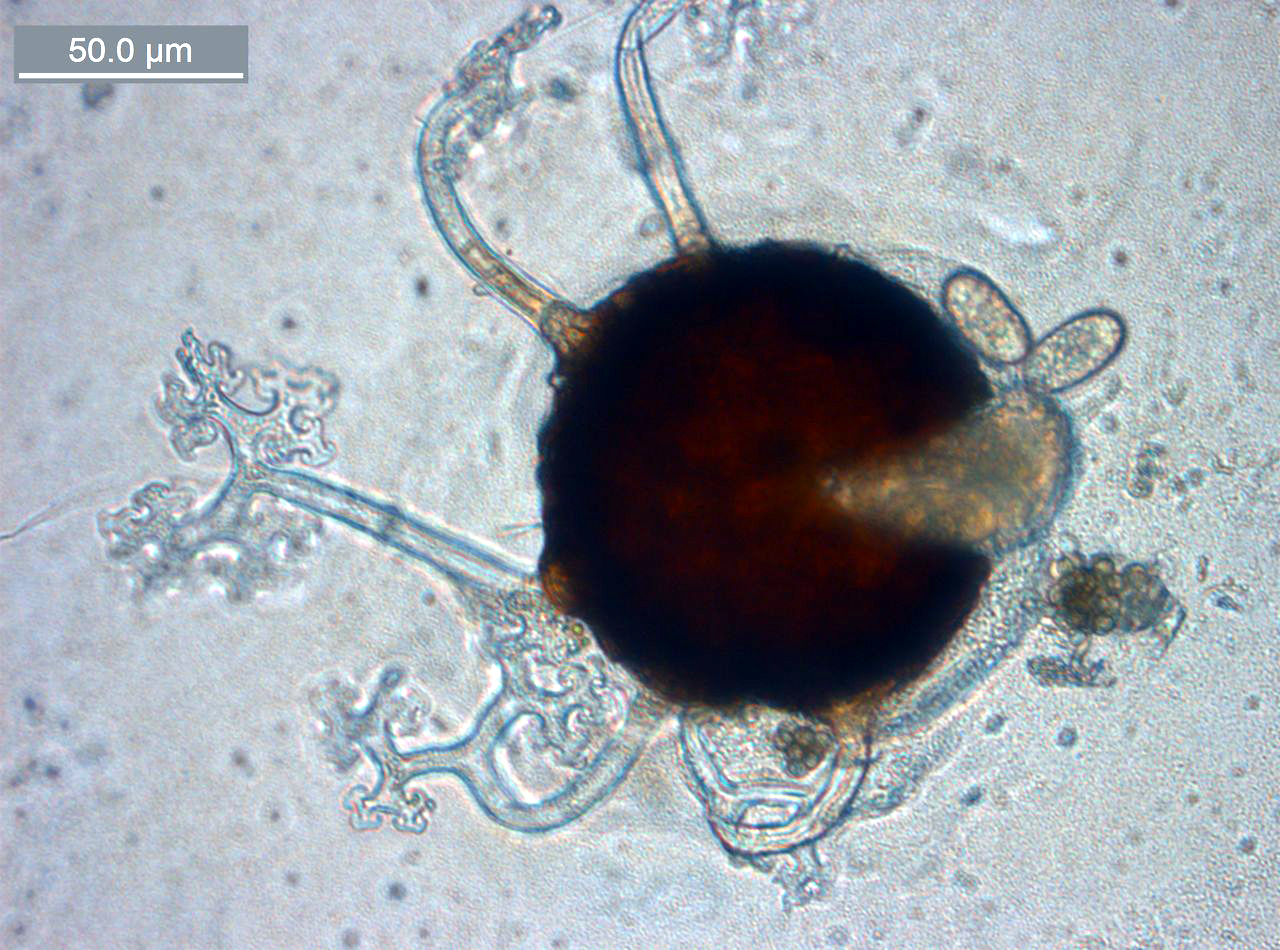
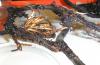
Cause Melampsoridium alni and M. hiratsukanum are present in Curry County, Oregon. M. hiratsukanum has been reported in British Columbia. An unidentified, and sparsely occurring, rust was observed on leaves along the McKenzie River in Oregon. It is not a very common or obvious problem around the region.
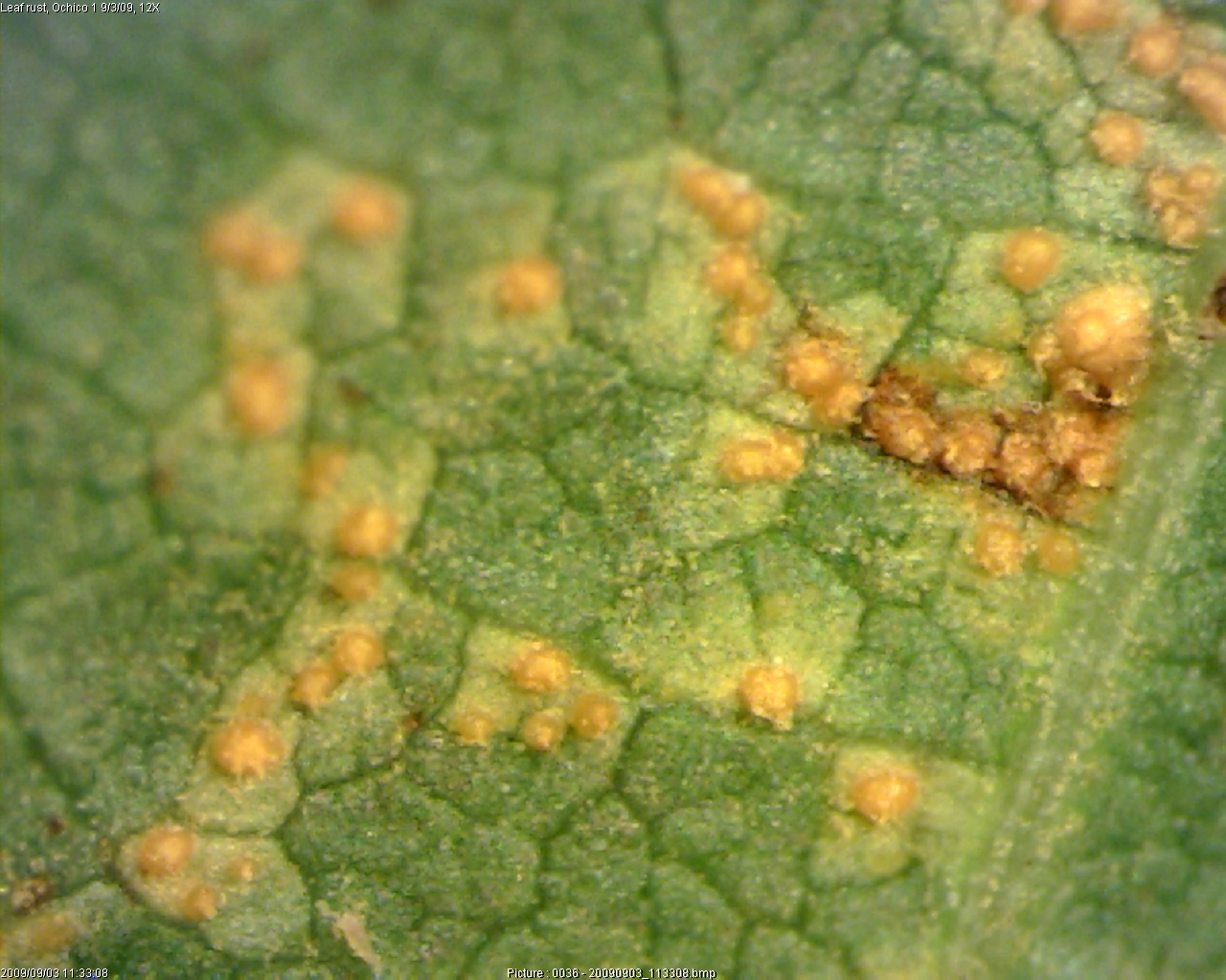
Melampsoridium sp. rust pustules on a red alder leaf.
Laura Sims, 2012.
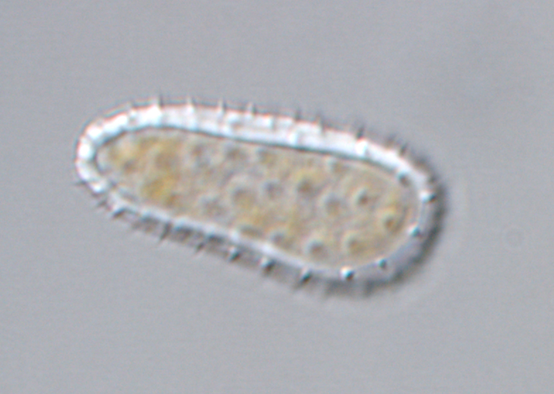
Melampsoridium sp. urediniospore from red alder rust pustule.
Laura Sims, 2012.
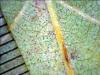
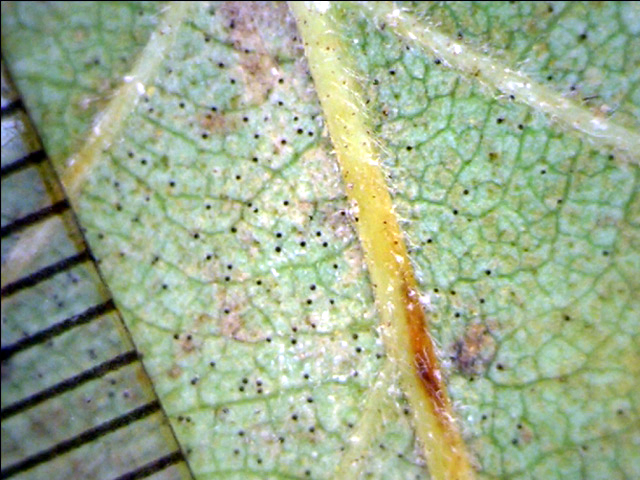
Cause Erysiphe aggregata, Erysiphe penicillata (formerly Microsphaera penicillata), and Phyllactinia alnicola have been reported from the Pacific Northwest. Generally does not warrant control.
Underside of red alder leaf with powdery mildew where black dots are chasmothecia. (scale on the left side is in mm).
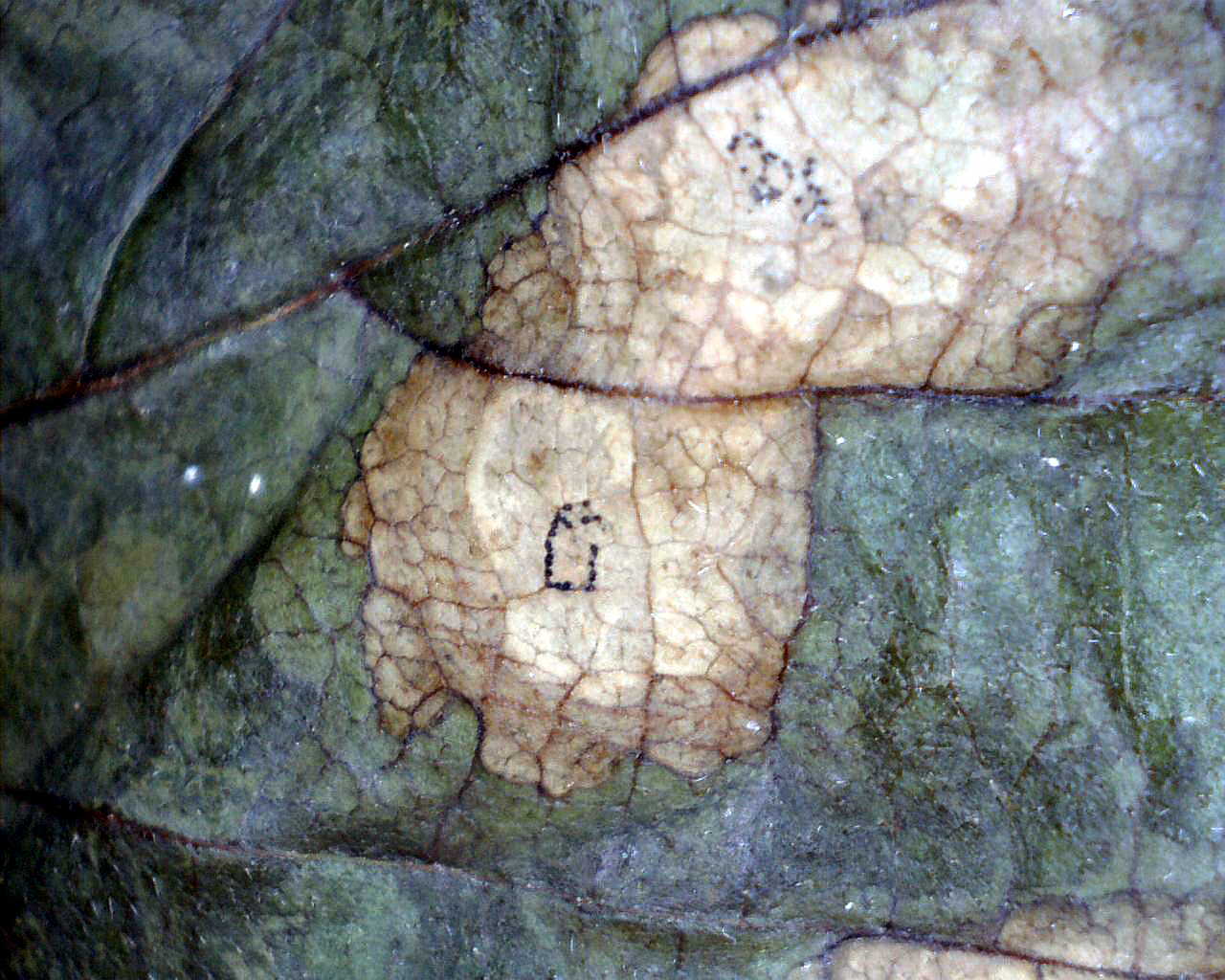
Cause Septoria alnifolia, a fungus that starts as a foliar pathogen and later causes severe stem cankers in alder nursery seedlings. Infected nursery seedlings with cankers must be culled prior to planting to prevent mortality and stem breakage.
Leaf spots on red alder have black pycnidia and can coalesce on leaf surfaces.
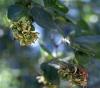
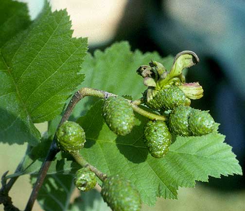
Note how the catkins are deformed by this fungus. Picture taken in the Blodgett Experimental Forest in California.
Photo by Everett Hansen, 1982.
Note how this catkin is deformed by this fungus. Picture taken in Idaho.
Photo by Everett Hansen, 1989.
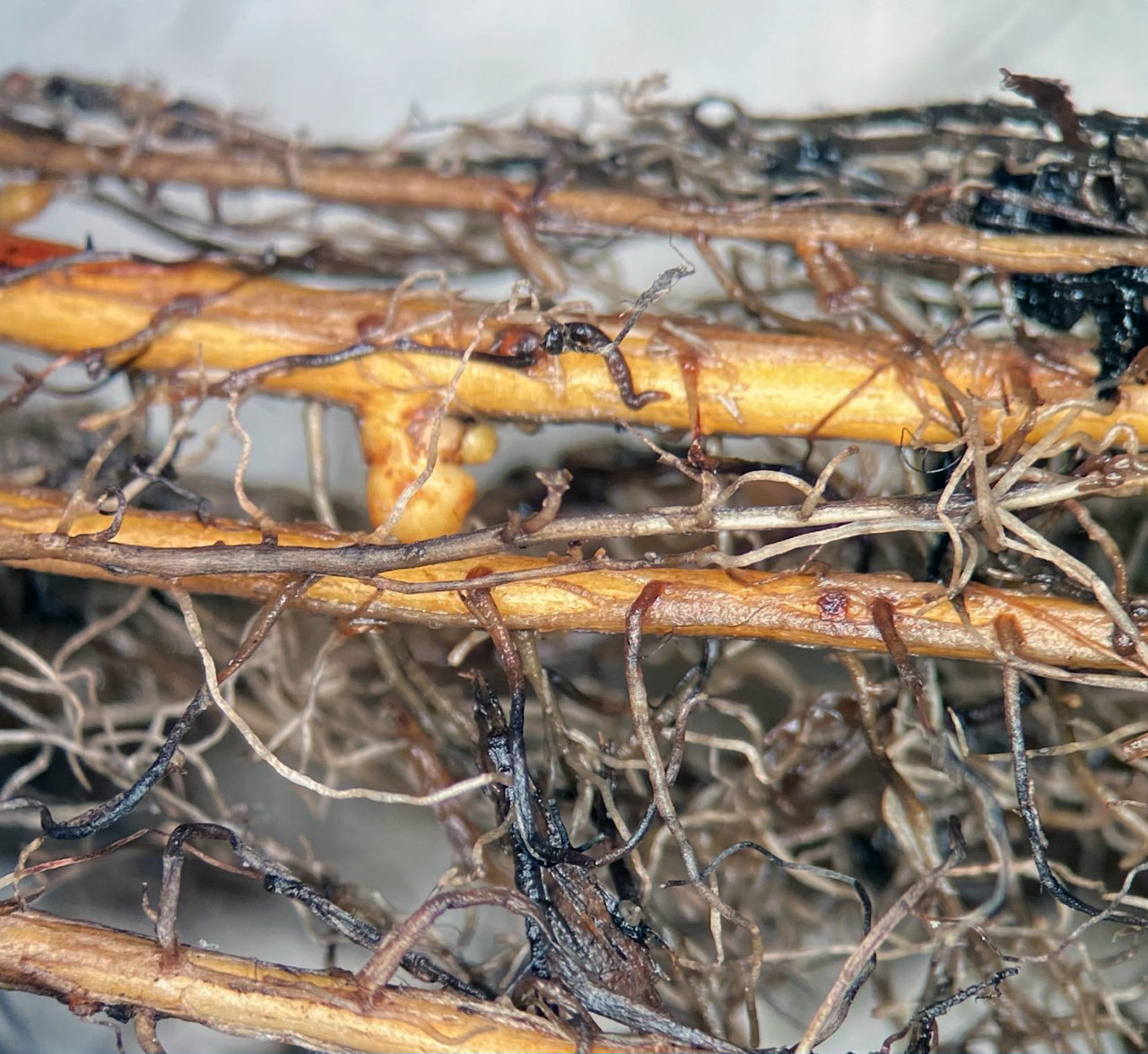
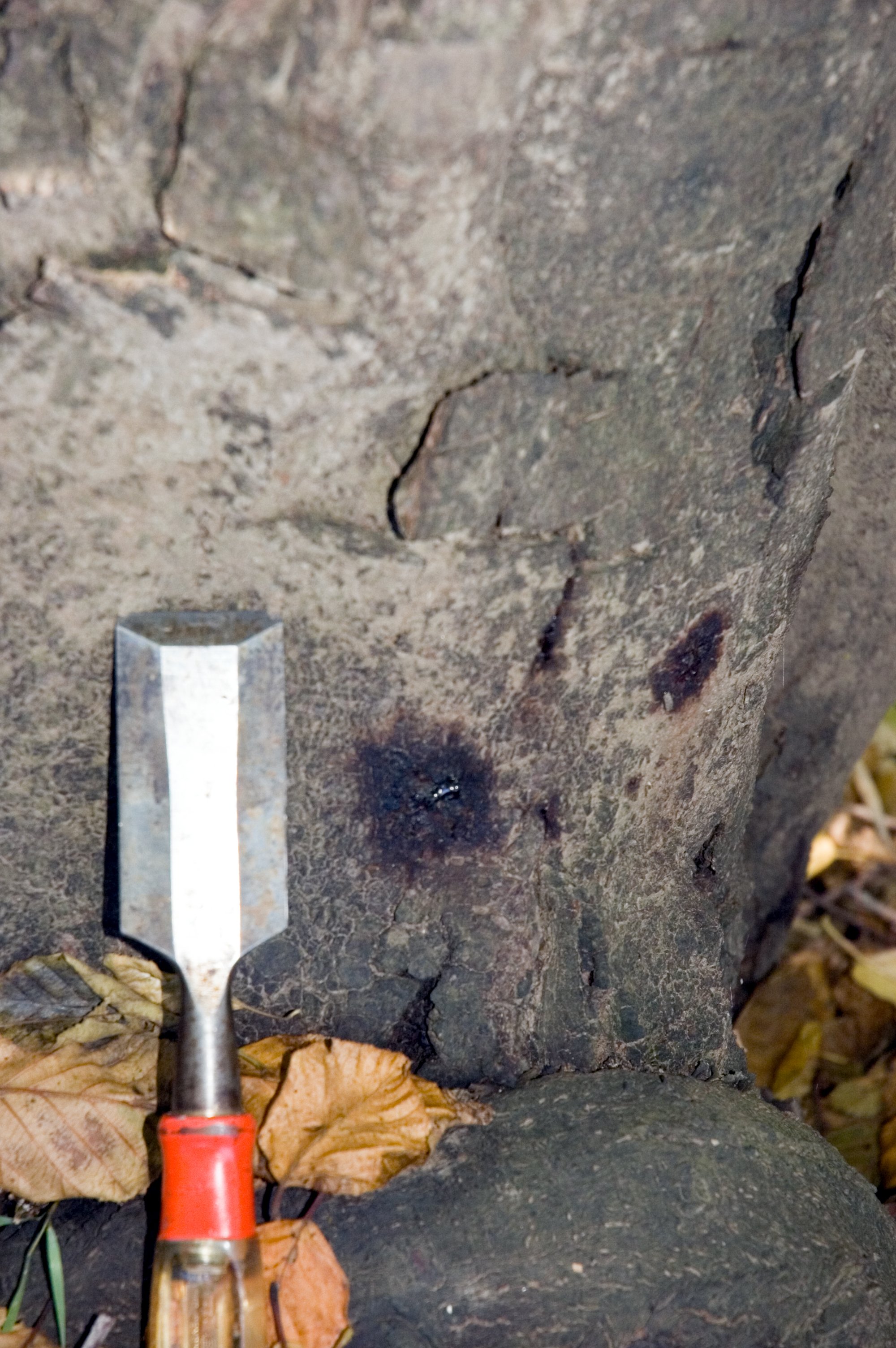
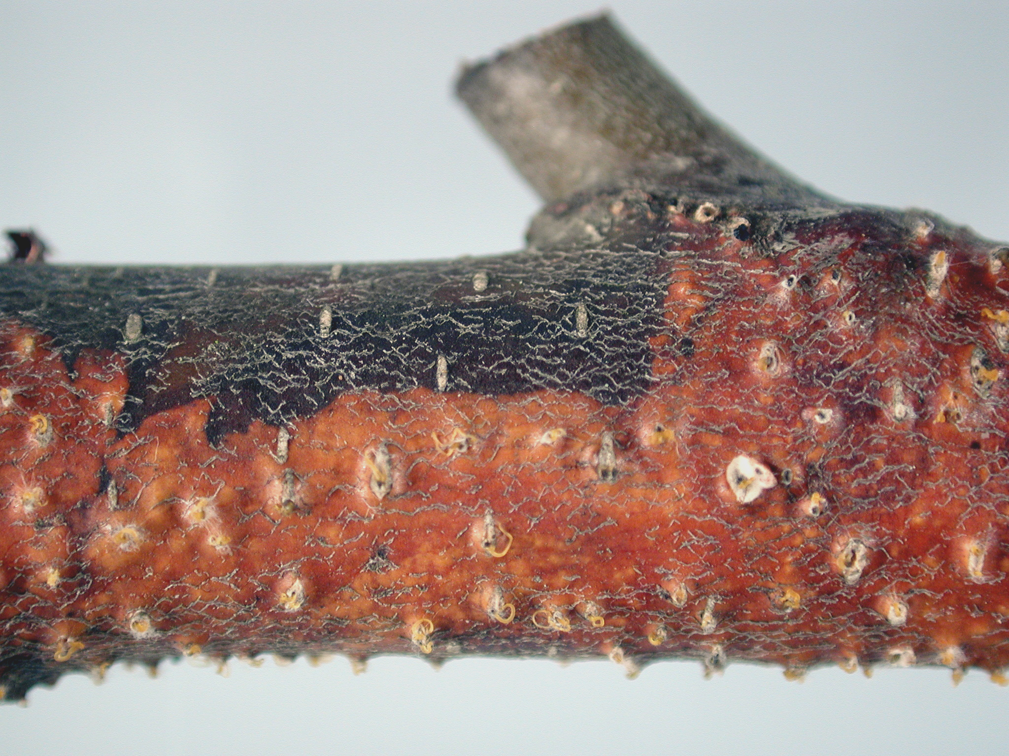
Cause There a number of fungi that can infect alder and result in stem decay and/or visible cankers. Few are economically important. Trunk rot is caused by Phellinusigniarius,which is reported to have some economic impact in timber, pulp, and recreation sites in British Columbia.
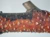
Diaporthe stem canker of red alder. Note the extruded coils of spores called cirri coming from pycnidia in the bark.
OSU Plant Clinic Image, 2013.
By L. Sims
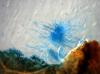
Cause Mycopappus alni (formerly Cercosporella alni)an ascomycete leaf pathogen forming filamentous conidia bunched in a mop-like mass. The conidial mass is held together by fungal stromatic tissue embedded in the leaf. These spore tufts are splash dispersed and readily dislodge from stromatic tissue by rain, dew or fog. Observed on red alders in Washington, Oregon, and on Vancouver Island, BC.
Filamentous conidia bunched in a mop-like mass. Blue color is from a stain used to view them under the microscope.