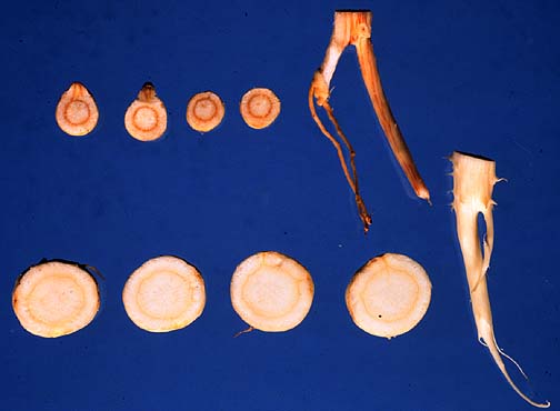
Note the smaller and discolored roots along the top. Circular root pieces have been cut in cross section.
Photo by Melodie Putnam, 1982.
By M. Putnam
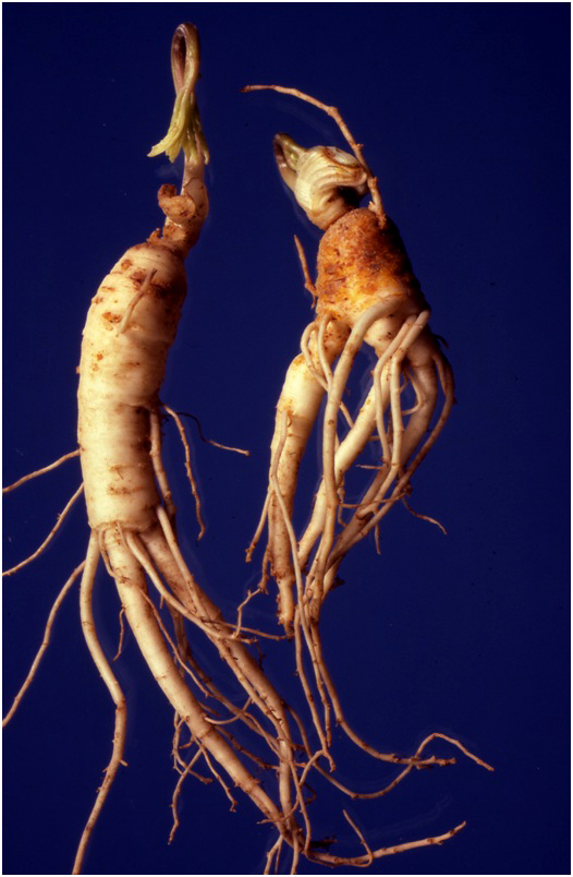
Four year-old roots showing the superficial “rusting” at the crown which is typical of this disease.
Melodie Putnam, 1983.
Cause The soilborne fungus-like microorganism Phytophthora cactorum. Spores are produced on infected foliage and in roots. Spores are spread via water splash, surface water runoff, and movement of equipment and workers through the beds. Spores also survive up to at least a year in decayed plant material. The fungus-like microorganism can infect foliage and move down into roots, although direct root infections are more common.
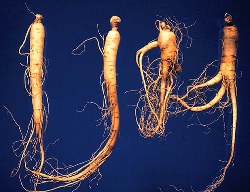
Various stages of this root rot.
Photo by Melodie Putnam, 1983.
Single sporangium on ginseng leaf.
Photo by Melodie Putnam, 1997
Foliar symptoms on ginseng of Phytophthora Rot.
Photo by Melodie Putnam, 1997
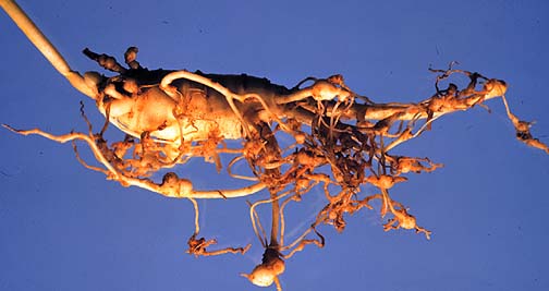
Cause The root-knot nematode, Meloidogyne hapla, does not penetrate to the interior of the root, but remain just beneath the surface. Galled roots are reduced in value.
Symptoms Leaf symptoms are typical of nonspecific stress such as generalized chlorosis, slight stunting, and premature reddening or yellowing.
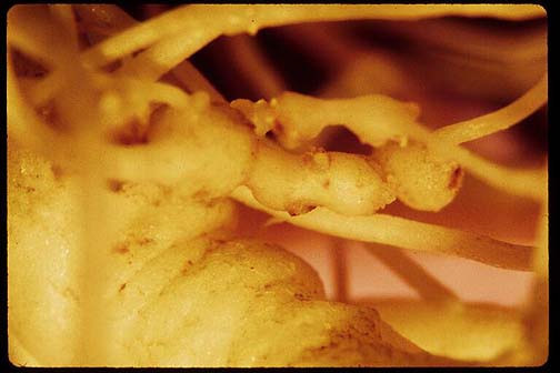
Note the swellings on the the smaller roots.
Photo by Melodie Putnam.
Note the swellings on these roots.
Kathy Merrifield
By M. Putnam
Cause Soilborne fungal and fungus-like microorganisms such as Rhizoctonia solani, Pythium sp., and Phytophthora sp. They are widespread in soil and can be very destructive under cool, moist conditions.
Phytophthora cactorum on ginseng.
Photo by Melodie Putnam, 1997.
Rhizoctonia solani on ginseng.
Photo by Melodie Putnam, 1983.
By M. Putnam
Note rust-colored lesions on the top two roots.
Photo by Melodie Putnam, 1983.
Note the few foliar symptoms associated with a root nearly completely rotted away, with only a hollow shell remaining.
Melodie Putnam, 1983.
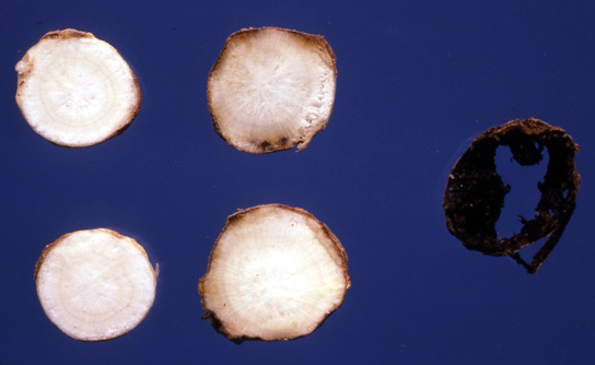
Very early stages of the disease shown in cross sections to the left and center, and a severely affected root cross-section at the right. The disease progresses to the interior of the root, eventually completely decaying it.
Melodie Putnam, 1983.
By M. Putnam
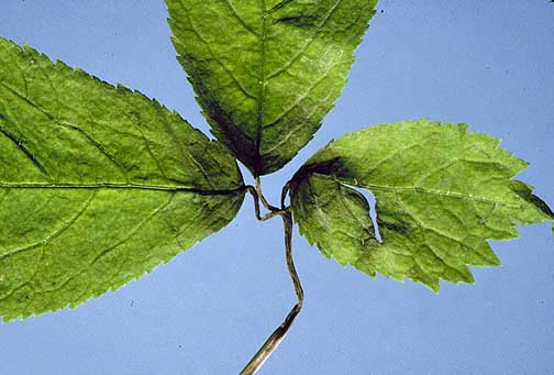
This picture shows the early stages of leaf and stem infection.
Melodie Putnam, 1982.
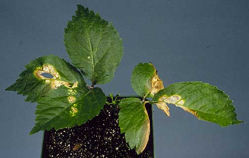
This picture shows later stages of leaf infection.
Photo by Melodie Putnam, 1983.
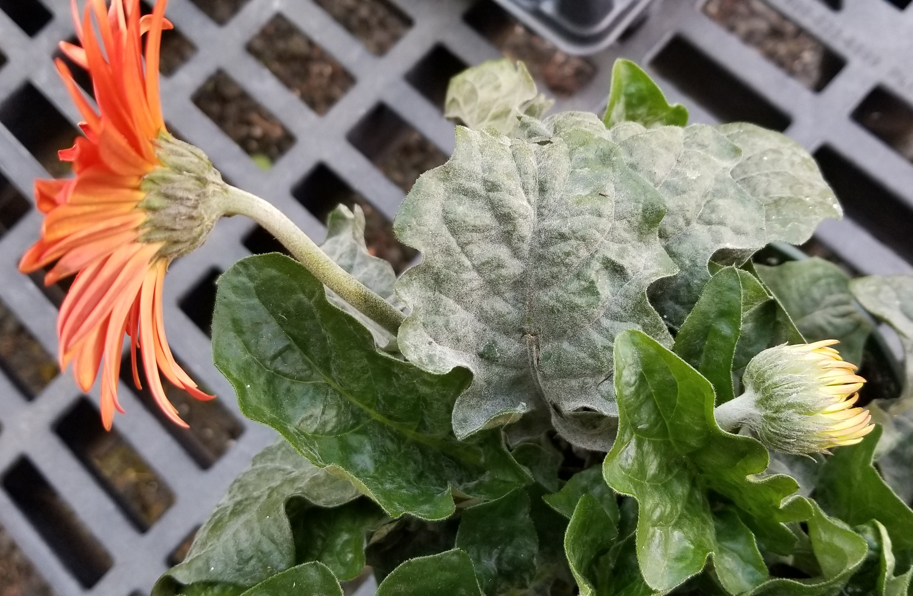
Cause Golovinomyces cichoracearum (formerly Erysiphe cichoracearum) is a fungus that is favored by moderate temperatures (68°F to 82°F) with alternating cool night and warm days and conditions that produce high humidity (80% to 90%) but dry leaves. A closed canopy and poor air circulation also promote disease development. It is a highly specialized pathogen that forms a close association with the host. Conditions that favor the host also favor the pathogen.
Powdery mildew can cover Gerbera leaves completely.
This old large format slide is labeled "White Mesh" which is likely referring to a disease caused by a virus now know as Pelargonium vein clearing virus (or maybe yellow net virus).
OSU Extension Plant Pathology collection, circa 1950s.
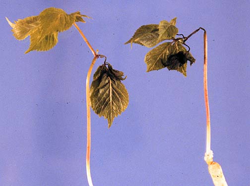
Cause Verticillium dahliae, a soilborne fungus that survives in soil indefinitely. It can spread in infested soil, container media, irrigation water, or in symptomless infected cuttings. The fungus grows into the xylem where it colonizes the plant through mycelial growth and conidial production. Fluid movement in the xylem passively transports the conidia. Once in the xylem, this fungus partially blocks water movement and produces toxins that result in wilt symptoms.
The plant on the left is wilting due to Verticillium wilt, while plant on right has leaf spot and stem rot.
Photo by Jay W. Pscheidt, 1992.
Wedge-shape areas on affected leaves turn yellow; then the entire leaf turns yellow and wilts.
OSU Extension Plant Pathology collection, circa 1950s, color glass slide plate.
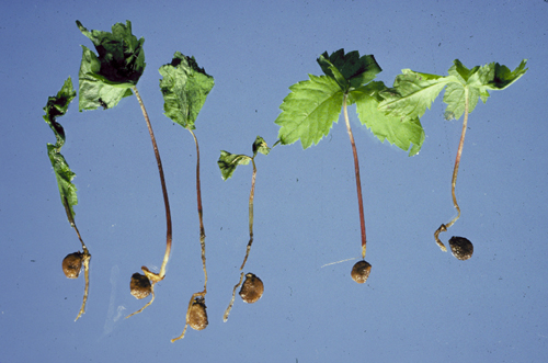
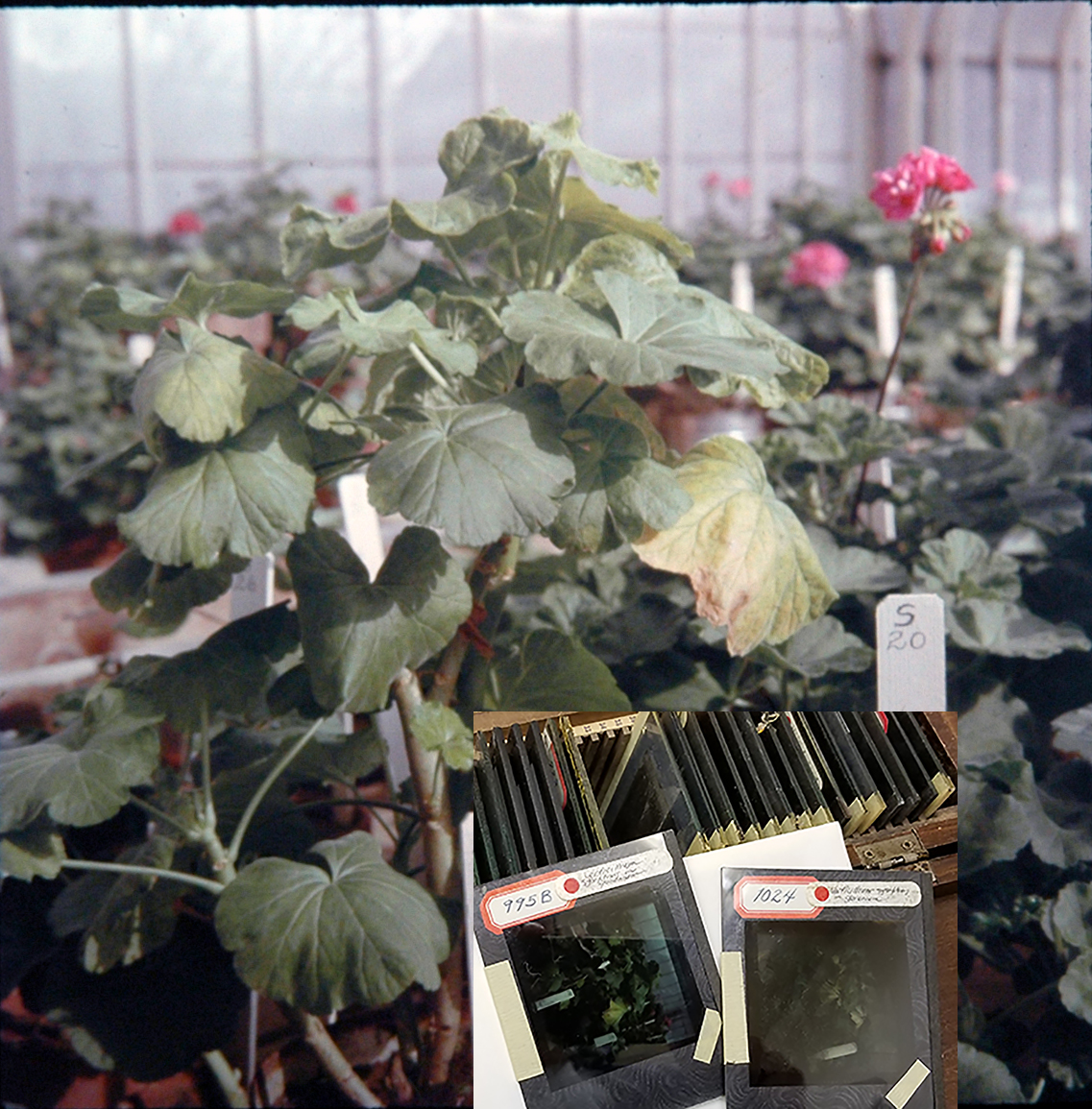
The stake in the pot reads sunburn, however, Verticillium wilt was found to be the cause of the wilting plants.
OSU Extension Plant Pathology collection, circa 1950s, from color glass slide plate.